
Ultrasound technique may help detect muscle changes earlier in SSc
Noninvasive tool ID'd increased muscle stiffness in early scleroderma, per study
Written by |
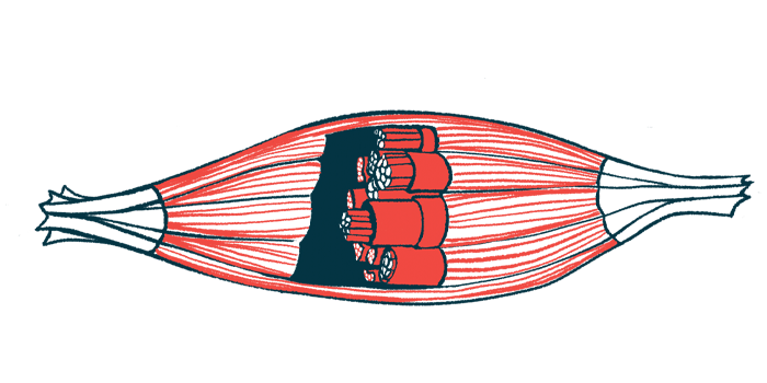




A noninvasive imaging tool called shear wave ultrasound elastography (SWE) helped detect increased muscle stiffness — specifically, a stiffer thigh muscle — in people with early systemic sclerosis (SSc), a study found.
These results suggest that SWE — a technique that measures the stiffness or elasticity of tissues — may detect muscle changes before they become apparent through routine clinical assessment. According to the researchers, “the clinical implications of these findings are potentially significant for the management of SSc patients.” In fact, the team noted that the technique may aid in quantifying muscle changes at an early disease stage.
“SWE offers the advantage of being a non-invasive, quantitative, and objective assessment tool, representing a substantial improvement over current diagnostic methods such as muscle biopsy, which is invasive, and MRI, which can be costly and less accessible,” the scientists wrote.
The study tested the tool in 30 people with early SSc and 30 age- and sex-matched healthy individuals, finding a stiffer thigh muscle among most patients despite them having no symptoms of SSc, also called scleroderma.
“Our findings support the potential utility of SWE as a complementary imaging tool for detecting early muscle involvement in SSc, “ the researchers wrote, noting, however, that it should not be used as a standalone test.
The study, “Evaluating the diagnostic value of shear wave elastography in early systemic sclerosis: a case-control study,” was published in the journal Scientific Reports.

SSc is caused by abnormal immune system activity that results in the thickening and hardening of the skin and connective tissues. Muscle involvement may cause stiffness, weakness, and impaired contraction, but early changes can be difficult to detect with accessible and practical tools.
SSc patients in study had no signs of muscle weakness
In other neuromuscular conditions, SWE has been used to provide an objective, quantitative measure of tissue stiffness. It works by analyzing how fast shear waves travel. However, little is known about muscle changes in people with early SSc without clinical signs of muscle weakness.
Now, researchers in Thailand investigated whether the technique could identify increased muscle stiffness at this early stage. Their study involved slightly more men than women, and the mean age at onset among the participants with early SSc was 50.1 years. As for disease type, most patients (77%) had diffuse SSc; the others were diagnosed with limited SSc.
The main symptom, affecting slightly more than 60%, was Raynaud’s phenomenon, a condition in which the fingers and toes feel numb, prickly, and frigid in response to cold temperatures or stress. Nearly half of patients had edematous skin, or tissue swelling right under the skin.
Using SWE, the researchers measured stiffness in several muscles of the thigh. People with SSc had significantly higher muscle stiffness in the left rectus femoris, located in the front of the thigh, than healthy controls. The other muscles assessed also tended to be stiffer in the SSc group, but the differences were not statistically significant.
Given that none of the patients in the study showed signs of clinical muscle weakness, the researchers noted that these findings may imply “that muscle can be changed stiffer before clinical symptoms manifest.”

Ultrasound tool suggested as complementary imaging method
The team also assessed how well muscle stiffness measurements could distinguish people with SSc from healthy controls. This was done using a statistical measure called the area under the curve (AUC). An AUC of 0.5 indicates that a test performs no better than chance, while a value of 1 represents perfect discrimination.
For the left rectus femoris, stiffness measured along the length of the muscle showed modest ability to distinguish the two groups, with an AUC of 0.67. Using a cutoff of 2.08 meters per second, the measurement correctly identified about 67% of affected participants and 70% of healthy controls. By comparison, stiffness measured across the muscle showed poor ability to distinguish between the groups.
The researchers noted that “the causes of … muscle involvement were not explored in this study, but could be multifactorial.” The team suggested scarring, muscle disease, restricted motion, or “steroid and other medical treatment” as potential factors, adding that “future study is needed.”
Overall, however, the scientists emphasized that this ultrasound technique should right now be viewed as a complementary imaging method rather than a standalone test.
Among the study’s limitations, the team noted, is its relatively small sample — which, they say, may explain why only the left rectus femoris showed statistically significant differences. Also noted was the lack of muscle biopsy to confirm that the increased stiffness represented actual muscle disease.
The researchers said future studies should compare SSc patients with or without proven muscle involvement, which would help “determine if SWE can differentiate not only between healthy and diseased states but also between different stages or severities of muscle involvement within the SSc population.”

Kathleen Edmond
I am female. My Scleroderma CREST experience age 10years.
First symptoms alongside fatigue was Raynaud's Disease. 1959. Scotland. Tests showed nothing! Hospitalised frequently. Diagnosed age 17 and was put on a clinical trial and given PENICILAMINE D, an immunosuppressant. By a Dr John Thompson in The Western hospital in Glasgow. He wrote medical papers about my problems. I should’ve mentioned that by the time I was 14 I had severe muscle wastage. I could hardly turn my head to either side. My hands were ‘clawed’, arms - difficulty tryingI to raise my arms above my head, elbows at a 90 degree angle couldn’t straighten, back - could hardly bend over, legs - knees could hardly reach a 90 degree angle. I will be 76 years old shortly and many of these issues, though a lot better since menopause, I still have problems with. My lungs - small airways disease, small in size, small input and output breathing. Heart - leaking valve, high BP, taking BP meds and Metoprolol. Arteries and veins damaged and severely shrunk. Can only be Canulated using ultrasound. Arteries in groin difficult pierce for angiograms. 2 heart attacks, 3 stents. 2 brain aneurysms. Sixth nerve palsy suspected stroke. Eseophagitis damage re severe reflux, poor motility. Difficulty swallowing. Voice box damage. GI disease. Poor absorption of nutrients and minerals. Iron, B12, Folate deficiencies. Suspected EDS and vEDS. Sjorgens disease affected eyes. Nose , sweat glands, gums, teeth. Vagina- so shrunk that it is difficult to have a ‘Smear test’ done. Affecting sexual health. I live in Australia now (warmer than Scotland). Hope this helps xx