Low Oxygen in Muscles Linked to Poor Exercise Capacity in SSc Patients
Written by |
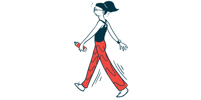


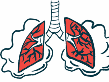
Poor oxygenation in skeletal muscles — those used in voluntary movements — contributes to impaired exercise capacity in people with scleroderma, a study suggests.
The study, “Reduced exercise capacity in patients with systemic sclerosis is associated with lower peak tissue oxygen extraction: a cardiovascular magnetic resonance-augmented cardiopulmonary exercise study,” was published in the Journal of Cardiovascular Magnetic Resonance.
Lung disease, heart muscle disease, anemia (low number of red blood cells), and pulmonary arterial hypertension (PAH) — a disease characterized by the narrowing of the small blood vessels, called pulmonary arteries, that transport blood through the lungs — are factors known to contribute to a limited ability to endure exercise, called exercise intolerance, in people with scleroderma.
Poor oxygenation in skeletal muscles contributes to the muscles’ poor performance. However, whether problems with skeletal muscles also contribute to exercise intolerance in scleroderma, also called systemic sclerosis (SSc), remains unclear.

To answer this, a team led by researchers at the University College London used a technique called cardiovascular magnetic resonance-augmented cardiopulmonary exercise testing (CMR-CPET) to assess exercise capacity by measuring simultaneously oxygen consumption and cardiac output (the amount of blood pumped out of the heart in one minute).
In total, the study (NCT04729777) included 60 participants — 15 patients with PAH-associated SSc (mean age 59.7); 15 SSc patients without PAH (52.5 years); 15 healthy participants, serving as controls (51.3 years); and a further group of 15 patients with nonconnective tissue disease (NC)-PAH, mean age 51.1. Participants with NC-PAH had either idiopathic (no known cause) pulmonary arterial hypertension or chronic thromboembolic pulmonary arterial hypertension.
In each group of SSc patients, with or without PAH, 14 patients had limited cutaneous SSc, and the other had diffuse cutaneous SSc.
Participants performed exercise on a supine CMR-compatible cycle ergometer, lying horizontally with the face and torso facing up. In the first minute, they had no resistance. Thereafter, the exercise was split in two-minute stages. In each stage, the workload increased every 30 seconds.
This was maintained until exhaustion, defined as an inability to maintain cadence or a verbal indication from the participant. Exercise was stopped and followed by a 15-minute recovery period with monitoring of vital signs.
For all participants, parameters were assessed at rest and at peak exercise. Additional collected parameters included T1 and T2 maps — MRI techniques that enable instant detection of heart abnormalities — hemoglobin levels, lung function, six-minute walk test (6MWT) scores, and cardiac catheterization.
Hemoglobin is a protein that carries oxygen in red blood cells; 6MWT, a measure of lung function, measures the distance an individual is able to walk over six minutes on a hard, flat surface; and cardiac catheterization requires the insertion of a very small, hollow tube into a blood vessel in the groin, arm, or neck for cardiac testing.
Lung function, measured by the 6MWT as well as by diffusion lung capacity for carbon monoxide (DLCO) — a parameter that assesses the ability of the lungs to transfer oxygen from the inhaled air to the blood — was significantly worse in SSc-PAH compared with NC-PAH. Further, DLCO was also lower in patients with SSc-PAH than in those with SSc only.
Results showed that all patients had significantly reduced peak oxygen consumption compared with healthy controls.
SSc-PAH patients had the lowest peak workload and shortest exercise duration. Compared with controls, those with NC-PAH had a lower peak workload.
Peak cardiac output was significantly reduced in both SSc-PAH and NC-PH patients compared with healthy controls and participants with SSc.
High hemoglobin levels were linked with higher peak oxygen content. Also, a higher myocardial T1 — a measure of heart muscle damage — was associated with less blood pumped out from the heart (divided by the body surface area).
Overall, SSC patients with or without PAH have a reduced peak oxygen consumption, suggesting “that tissue oxygen extraction is an important determinant of exercise intolerance in SSc and could be used as a biomarker of disease and response to therapy,” the team concluded.