
Levels of Specific Cytokines Linked to Clinical Manifestations of Scleroderma, Study Reports
Written by |


Levels of certain important signaling molecules called cytokines are associated with different clinical manifestations in patients with scleroderma, a study suggests.
The study, “Different profile of cytokine production in patients with systemic sclerosis and association with clinical manifestations,” was published in the journal Immunology Letters.
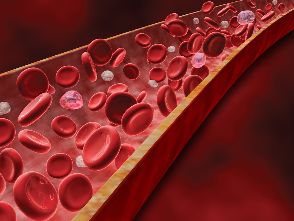
Cytokines are important molecules that can induce tissue damage, recruit additional inflammatory cells, and promote fibrosis, the thickening or scarring of connective tissue.
In this study, researchers investigated how the levels of specific cytokines in the blood of scleroderma patients correlate with their clinical manifestations. They obtained blood samples from 56 scleroderma patients — 53 women and three men — with a mean age of 45.1 years. The same number of unrelated age- and gender-matched healthy individuals were analyzed as controls.
Unfortunately, the levels of several cytokines — interleukin (IL)-2, IL-4, IL-10, IL-6, and tumor necrosis factor (TNF) — were too low to be measured.
Levels of the cytokine IL-17A — previously shown to play a role in the inflammation of scleroderma — were found to be higher in scleroderma patients than in healthy controls. The difference, however, was not statistically significant.
Comparing patients showing organ involvement with those without it, only patients with lung fibrosis showed a significant increase of IL-17A levels.
“Since the levels of most cytokines were undetectable in the serum, we decided to evaluate the production of these cytokines by PBMCs [peripheral blood mononuclear cells] from scleroderma patients and healthy individuals,” the researchers wrote.
PBMC is a general designation that denotes any blood cell with a round nuclei, and includes immune cells like monocytes, lymphocytes, and macrophages.
In unstimulated PBMCs (without any external stimuli), the base levels of TNF, IL-10, IL- 2 and IL-6 were already higher in scleroderma patients than in healthy controls, suggesting that PBMCs in patients produce higher amounts of cytokines.
After dual stimulation — in this case with anti-CD3 and anti-CD28 molecules — PBMCs from scleroderma patients had lower concentrations of TNF, IL-10, and IL- 2 than controls. Whether this decrease is linked to a lower cytokine production per cell or to a low number of cells capable of producing cytokines among PBMCs remains unknown.
Looking at how the levels of cytokines correlate with clinical manifestations of scleroderma, researchers found that in unstimulated PBMCs, IL-2 concentration was higher in patients with esophageal motility dysfunction. Likewise, the cytokine IL-10 showed a significant positive correlation with a modified Rodnan score, a measure of skin thickness used as a key outcome parameter in clinical trials of scleroderma.
After stimulation, significantly higher levels of IL-2 and IL-4 were detected in scleroderma patients with lung fibrosis; and higher IL-10 and IL-4 in those with digital ulcers.
“In conclusion, we identified substantial differences in cytokine production in SSc [scleroderma] patients compared to healthy subjects. This altered cytokine production was associated with cutaneous and involvement of internal organs,” the researchers wrote.
The team suggests that the findings “support the view that chronic cellular activation occurs in SSc, which leads to persistent release of cytokines involved in its pathogenesis.”


Leave a comment
Fill in the required fields to post. Your email address will not be published.