
Free Light Chains in Blood and Urine May Aid in Diagnosis, Mark Activity
Written by |


The amount of free light chain (FLC) molecules in blood and urine correspond to the severity of scleroderma, adding to the evidence supporting these molecules as biomarkers for early diagnosis and disease activity.
The pilot study detailing this finding, “Serum and urine free light chains measurements in patients with systemic sclerosis: novel biomarkers for disease activity,” was published in the journal Clinical & Experimental Immunology.
Free light chain molecules are made during the production of antibodies, and are considered to be biomarkers for B-cell activity. B-cells, a part of the immune system, tend to become self-reactive in scleroderma, or systemic sclerosis (SSc), meaning that they target the body’s own tissues, causing inflammation.
Activated B-cells produce antibodies — molecules that “flag” cells and other biological molecules for the immune response — called immunoglobulins. Immunoglobulins are made of light and heavy chains (two of each). The two light chains on each immunoglobulin will both be of a certain variant, called kappa and lambda.
Free light chains are the excess light chains made by activated B-cells, and released into the blood. Here, they tend to be rapidly removed from the body by the kidneys, potentially making them direct biomarkers of B-cell activity.
Despite their association with B-cells and reports that they also associate with scleroderma activity and severity, as well as with scleroderma-related interstitial lung disease, few studies have evaluated their levels in this disease and none has examined their presence in urine.
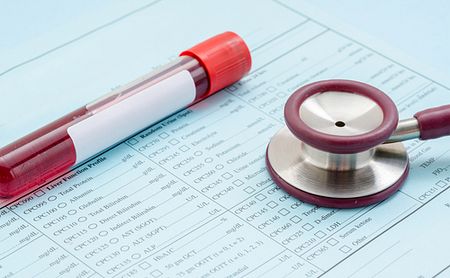
To address this knowledge gap, researchers in Italy analyzed FLC levels in the blood and urine of 72 adult SSc patients (median age, 55) and 30 healthy controls, seeking to correlate these levels with disease severity and activity.
People with scleroderma showed significantly higher amounts of both kappa and lambda FLCs in their blood than did the control participants, with kappa FLC levels generally higher than those of lambda.
Likewise, urine levels of kappa FLCs were significantly higher in patients than in healthy controls, but no such significant difference between the two groups was seen in lambda FLCs.
Greater levels of both FLCs tended to correspond to higher measures of inflammation, as assayed by C-reactive protein levels, and a faster erythrocyte sedimentation rate — a test that measures how fast red blood cells settle at the bottom of a test tube. They also correlated with more disease activity and severity, as measured using the disease activity index (DAI) and the disease severity scale (DSS).
DAI scores rose with urinary kappa FLCs, as SSc patients with more of these had statistically higher DAI scores than those whose kappa FLC levels remained below 15.1 mg/L.
“We suggest that FLCs could be employed as reliable and useful potential biomarker of early diagnosis and to follow disease activity,” the team wrote, “leading to an optimization of SSc patients clinical management.”
Study limitations mentioned by the investigators included its relatively small sample size and the fact that enrolled patients had a long disease duration (median of 11 years).


Leave a comment
Fill in the required fields to post. Your email address will not be published.